Workshop on Neuroimaging

Written by Yingchen

Edited by Xin Chen, Sasinthiran and Keshiniy

20160305191608
Figure 1: Members of NUS Neuroscience Student Interest Group at Clinical Imaging Research Centre

On the 22nd of February 2016, members of NUS Neuroscience Student Interest group attended a workshop on Neuroimaging hosted by A*STAR-NUS Clinical Imaging Research Centre (CIRC).

The Workshop started with an introductory lecture by Dr John Totman, Head of Imaging Operations at CIRC. He recounted the history behind various imaging techniques such as Ultrasound, X-Ray, CT, PET and MRI, and gave a brief overview on the science behind these techniques, their limitations and common uses, for example, in the field of nuclear medicine.

One of the most common imaging techniques, ultrasound makes use of the Doppler Effect.  Ultrasound waves are emitted from the probe, which then detects the reflection of those waves off anatomical structures to construct an image of the structure; similar to echolocation used by dolphins and bats. Ultrasound is not commonly used in neuroimaging because sound waves cannot penetrate the skull.

Another commonly used technique, X-ray works on the principle that structures in the body attenuate x-rays that are projected on one side of the body such that the rays emerging from the other side expose a sheet of film to produce an image reflecting the body structures. However, the images produced are two-dimensional and thus may miss out certain structural details. As a result, multiple images at different sections of the body are required for a more accurate representation of the body structures.

A variant of X-ray imaging is Computerized Tomography (CT). It uses a rotating x-ray emitter and detector to provide a 3D reconstruction of anatomical structures. It is commonly used in neuroimaging to provide the structural information that aids in medical practices, such as detecting tumours and aneurysms. The grey and white matter of the brain can be distinguished clearly on CT images.

Another technique, Positron Emission Tomography (PET) requires an ingestible tracer consisting of glucose in conjugation with radioactive fluorine, which is prepared in the expensive cyclotron reactor by accelerating and colliding sub-atomic particles. Other positron-emitting radionuclides such as oxygen might also be used depending on how long the effect is required to last. When the radioactive substance decays, positrons are released which collide with the electrons released by the PET scanner, producing two gamma rays which scatter in exactly opposite directions. The PET scanner detects these two gamma rays and computes the average distance at which the rays originated to indicate the location of the anatomical structure. PET can be used for functional as well as anatomical imaging. The principle behind anatomical imaging is that cancer cells take up glucose rapidly, but fludeoxyglucose (FDG) cannot be easily metabolised and thus accumulate in these cells and can be detected.

SPECT, which stands for Single-photon Emission Computerized Tomography, is a combination of PET and CT. PET provides metabolic information while CT provides structural anatomical landmarks such as bones to give relevance to the metabolic activity reported by PET scans.

After Dr Totman’s lecture, Ms Caroline Wong, a Research Officer at CIRC, introduced us to Functional Magnetic Resonance Imaging (fMRI), which is the most commonly employed imaging tool for functional studies in neuroscience research. fMRI provides both structural and functional information of the brain. The basic unit of fMRI images is the voxel, which is a 3D pixel with modifiable size and length. Smaller the voxel, greater is the time required to acquire it, but more detailed the image would be, which is analogous to having thinner slices: many voxels make up a slice, and many slices make up the volume.

The science behind MRI is that protons have different spins in random directions, which align when placed in a magnetic field. When the MRI scanner emits radio frequency pulses, the protons are tilted out of alignment from each other. When the radio frequency pulses stop, the protons lose energy and return to their baseline aligned state, thereby releasing electromagnetic waves that are detected by the scanner.

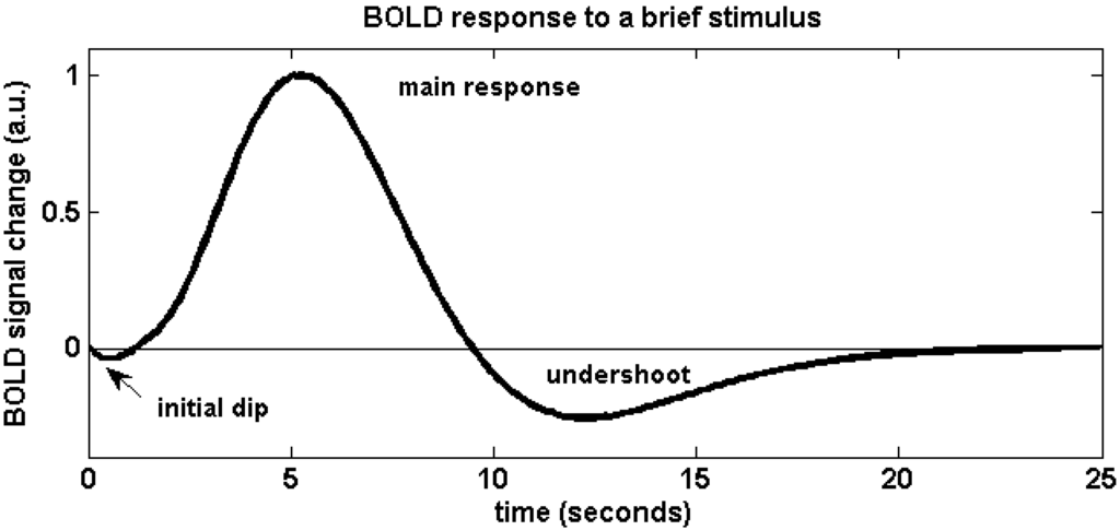
In fMRI practice, active areas of the brain receive more oxygenated blood flow due to the dilation of local cerebral blood vessels. The protons in oxygenated haemoglobin and deoxygenated haemoglobin have different rates at which they return to their baseline aligned state. Moreover, oxygenated blood is diamagnetic, does not distort the surrounding magnetic field and thus there is no signal loss. In contrast, deoxygenated blood is paramagnetic, distorts the surrounding magnetic field and thus there is signal loss. These differences are used to highlight brain areas that are active during a task, since active areas are marked by a higher level of oxygenated blood flow. A more detailed relation is displayed by Figure 2, the hemodynamic response function profile.

materials-04-01941-g001-1024
Figure 2: Hemodynamic Response Function Profile (MDPI.com)

In Figure 2, the initial dip is due to blood oxygen taken in by active brain areas from surrounding blood vessels. The rapid rise is due to overcompensation of blood flow due to dilation of blood vessels, which usually lasts for around 4-8 secs, but the exact time period depends on the brain area. The post-stimulus undershoot is due to elastic recoil of expanded blood vessels. Meanwhile, voxel colour changes with the time course of the curve.

Typical fMRI task designs include the block design, which consists of periods of rest between periods of activity; the slow event-related (ER) design, which has been phased out as it is inefficient; the rapid counterbalanced ER design, which is the fastest; and the mixed design, which consists of both block and ER. One difficulty with fMRI is that it requires minimal movement of non-task related areas of the body since movements may create “noises” that confound the results. Another difficulty is that brain functions are not localised to particular areas; meanwhile, the same area may be involved in many different functions. Notably, synchronisation between multiple brain areas that process the same cognitive properties have been seen to be out of synchrony in patients with neuropsychological disorders such as schizophrenia and bipolar disorder. Thus, careful interpretation of neuroimaging results is necessary.

After the presentations, we were invited to visit the fMRI laboratory, and witnessed several interesting phenomena such as the strong force of attraction between metal objects and the fMRI scanner, and the fact that conductors would fall due to gravity when suspended, albeit at a slower than expected rate in the fMRI scanner chamber due to electromagnetic induction. Towards the end of the workshop, Ms Caroline Wong also indicated that Dr Qiu Anqi from NUS Computational Functional Anatomy Lab was looking for research assistants. Details can be found in this link: http://www.bioeng.nus.edu.sg/cfa/.

Within the two-hour session, the workshop provided us with an essential foundation for appreciating the various neuroimaging techniques used in neuroscience research and clinical practice. We would like to thank the Clinical Imaging Research Centre (CIRC) for accommodating our group and conducting this workshop.

Reference:

“MDPI Open Access Publishing”, MDPI AG, 1996-2016. Accessed 7th March 2016. (http://www.mdpi.com/materials/materials-04-01941/article_deploy/html/images/materials-04-01941-g001-1024.png.)

Seminar on Cellular Neuroscience

Written by Sasinthiran

Edited by Xin Chen, Yingchen and Keshiniy

potassium_channels-264px.gif
Image: Overhead view of a voltage-dependent potassium ion channel shows four red-tipped “paddles” that open and close in response to positive and negative charges. (https://www.bnl.gov/bnlweb/history/nobel/nobel_03.asp)

On the 12th of February, members of NUS Neuroscience Student Interest group convened for a seminar on Cellular Neuroscience, as part of the fortnightly seminar series hosted by group members to explore different topics in neuroscience. The seminar was hosted by Enos Goo (Year 3, Life Sciences).

The seminar started off with a presentation on the basic principles of electrophysiology and the electrochemical basis of the membrane potential that is key for the generation of Action Potentials in neurons. Enos then touched on the remarkable diversity of individual neurons in terms of the vast variations in ion channels that confer unique properties when expressed in neurons. Differential expression and distribution of such ion channels allow for unique modulation of the membrane potentials of neurons and thus contribute to their diversity.

Next, Enos shared about specific types of ion currents such as the dendritic A-type current, and the dendritic H current which confer different electrophysiological properties to neurons through the action of specific ion channels. For example, it was noted that thalamic spindles observed during sleep are a result of an interaction between a calcium ion current and an inward pacemaker dendritic H current in neuronal populations at the thalamus. This property allows for the generation of rhythmic bursts of action potentials  that allows for a reduction in relaying of sensory input from sense organs to higher cortical areas for processing, since the thalamus functions as a relay centre for relaying such information (save for that of olfactory input).

It was noted that while science takes a reductionist approach in understanding the mechanisms of action in biological systems, a more global perspective yields greater insight into the more complex emergent properties of the components of such a biological system working together. An analogy was drawn to to our human society which is composed of numerous unique individuals, with not one of us being indispensable to the functioning of society as a whole. Thus, studying how a population of diverse neurons work together in neural circuits to give rise to complex emergent properties such as consciousness, the notion of morality etc., warrants significant interest.

It was also shared that back-propagated action potentials can be recorded in dendrites (albeit with a slight delay) as they too possess voltage-gated channels to support the propagation of action potentials.

The second part of the seminar involved discussing the notion of Intrinsic plasticity which involves the dynamic modulation of the electrophysiological properties of a neuron which affects their excitability and the computation of spatial and temporal summation of input the neuron receives ( as opposed to synaptic plasticity which is implicated in learning and memory) and its implications in disorders of the central nervous system. It was noted that in aging, an enhanced after-hyperpolarisation is seen at the end of action potentials and that this has been attributed to aberrant functioning of potassium ion channels which usually return the neuron to its resting membrane potential at the end of an action potential. The aberrant functioning of such channels with age could be due to the accumulation of mutations throughout life.

In epilepsy, it was noted that there was an aberrant down-regulation of potassium ion current and an enhanced persistent sodium ion current, leading to an increased intrinsic excitability of the neurons and hence the rapid burst of action potentials characteristic of an epileptic episode. In neurodegenerative diseases such as Alzheimer’s Disease (AD) and Parkinson’s Disease (PD), changes in the electrophysiological properties of the neurons have been observed prior to neuronal death (e.g. changes in ion channel expression and distribution). Moreover, degeneration of the Septal nuclei in AD leads to reduced cholinergic input to the hippocampus. This leads to an increased M current (efflux of potassium ions) and an enhanced after-hyperpolarisation of hippocampal neurons following action potentials, thus leading to the suppression of the neurons which usually fire in rapid bursts. It is noteworthy that firing in rapid bursts is an essential trait for memory coding and consolidation.

The seminar concluded with an interactive discussion on a few key topics. First, we reviewed the role of astrocytes in the ‘tripartite synapse’ and acknowledged the paradigm shift in considering the role of glial cells in the nervous system from one that views them as passively contributing to the matrix that hold neurons together in the brain to the view that they have active modulatory effects on neuronal activity (e.g. tripartite synapse’).

Next, we discussed the use of optogenetics (transfection of genes coding for light-sensitive rhodopsin activated ion channels into neurons) and considered potential applications of this technology in treating depression for example by activating serotonergic neurons instead of relying on drugs that have side-effects. However, the limitation of the technology is that light has to be directed towards the population of neurons via an optic cable (invasive). We also briefly discussed the possible physiological mechanism through which populations of neurons in migratory birds an other animals can use the earth’s magnetic field patterns to guide their migrations. We considered the possibility of using AC magnetic fields to heat (via eddy currents) a nanoparticle fused to an ion channel as a means of controlling the closure of the inactivation gate. This allows for a non-invasive way to control the activation of specific neuronal populations (a greater degree of spatial resolution).

Finally, we discussed the fact that a single nucleotide substitution on the voltage-gated sodium ion channel gene in pufferfishes allow them to be resistant to their own toxin (tetrodotoxin) which paralyses the nervous system of those who come into contact with it by inactivating the voltage-gated sodium ion channels. thus no action potential can be generated as the membrane cannot be depolarised by an influx of sodium ions. It is notable that this discovery was made by a research team based at NUS, headed by Professor Soong Tuck Wah.

The slides used for the presentation during the seminar can be found here (courtesy of Enos Goo): Cellular Neuroscience presentaion slides [943273]

Seminar on Neurons, Glia and Neurophysiology

Written by Sasinthiran

Edited by Xin Chen, Yingchen and Keshiniy


On the 29th of January 2016, the NUS Neuroscience Student Interest Group conducted its first seminar titled ‘Neurons, Glia and Neurophysiology’, hosted by Sasinthiran (Year 3, Life Sciences).

Participants were provided reading material on the topic compiled from various sources two weeks prior to the seminar. At the start of the seminar, participants attempted to complete a quiz (18 questions) in groups of 3-4 members within 30 minutes.

_20160209_181639 [2344380].JPG

The quiz questions were then discussed in an interactive format whereby groups had to defend and justify their answers if they differed from that of other groups.

The discussion started off with a question on the permeability of the blood-brain barrier (BBB). It was first highlighted that the components of the BBB are the tight junctions between the capillary endothelial cells, the basal lamina of the capillary and the perivascular foot processes of astrocytes (a type of glial support cell). It was noted that water and water soluble agents were able to diffuse across via the tight junctions while charged species such as sodium ions were first transported by trans-membrane protein pumps/ channels into the endothelial cells of the blood capillaries and subsequently into the brain. Lipid soluble molecules are free to diffuse through the phospholipid bi-layer of the endothelial cell membranes while larger charged molecules such as glucose ( primary energy source of the brain) and amino acids are transported by by trans-membrane transport proteins. Other solutes may also be transported by receptor-mediated endocytosis when they bind to their specific receptors on the surface of the endothelial membrane, as well as adsorptive transcytosis. It was noted that due to tight junctions between the capillary endothelial cells, leukocytes a(white blood cells) and other immune cells may not be able to cross the BBB to a great extent and hence the brain relies on a local population of cells known as the microglia (not derived from neuroectodermal lineage but from immune cell lineage) which mediate local immune function (e.g. phagocytosis and secretion of inflammatory factors such as interleukins to attract other immune cells).

Next, the discussion focused on the profile of an action potential (AP). It was noted that the refractory period (decrease in membrane potential to a value more negative than the resting membrane potential) after the peak in voltage of an AP had two parts: the absolute refractory period (lasting 1 msec due to the closing of the inactivation gate of sodium ion channels) during which no  action potential can be generated followed by the relative refractory period during which a higher than usual stimulus is required to depolarize the membrane to threshold potential in order to trigger an action potential. It was also noted that the fall in membrane potential below that of resting potential because the voltage-gated potassium channels that opened at the peak of the AP were slow to close and hence allowed more potassium ions to diffuse out of the neuron, down its concentration gradient.

The discussion then looked at the factors affecting the speed of conduction along an axon and 4 factors were identified: higher temperature, wider diameter of the axon, increased myelination (insulation) and decreased length (between nodes of ranvier) all favor faster conduction. It was also noted that different classes of axons with different diameter and myelination properties exist in the body to serve their particular functions. for example, when someone hits his toes against a hard surface, the faster A-delta fibres that are myelinated conduct a sharp pain sensation while the slower unmyelinated C fibres conduct the slow, dull pain that follows.

Next, we reviewed the various gating mechanisms of ion channels: ligand-gated, mechanosensitive (e.g. stretch-activated), voltage-gated, photon-gated (responds to light) or even ungated. We briefly discussed the recent development of optogenetics in which genes coding for a light-sensitive receptor has been transferred and expressed in select populations of mice neurons to modulate their behavior when that region is exposed to light via an optic fiber cable.

Our discussion then moved on to define the terms that describe a cell membrane’s potential. Depolarisation represents a rise in membrane potential from a negative resting potential towards the potential of 0mV (same internal potential as outside of the cell). Repolarisation represents a drop in membrane potential from 0mV to a more negative potential. Hyperpolarisation refers to a drop in membrane potential to a value more negative than that of the resting potential.

Next, we had a brief review of the activity at the neuromusccular junction (NMJ) that results in the contraction of skeletal muscle fibres for movement. It was noted taht acetylcholine was the primary neurotransmitter released at the NMJ.

The discussion then moved on to briefly touch on the fact that groups of axonal tracts that run up and down the spinal cord are arranged in columns. It was noted that such tracts are called projections when observed in the brain.

We then proceeded to discuss the factors responsible for the establishment and maintenance of the action potential: the unequal distribution of ions inside and outside the neurons (sodium, chloride and calcium ions are concentrated outside the cell while the inside of the cell has a higher concentration of potassium ions and other large non-difussible anions that also contribute to the negative resting membrane potential), the action of the sodium-potassium ATPase pump which pumps out 3 sodium ions and pumps in 2 potassium ions against their concentration gradient (thereby resulting in the net loss of one positive ion) as well as the free diffusion (leakage) of potassium ions out of the cell through leaky potassium channels.

Next, the discussion focused on the role of glia in the nervous system. It was noted that there has been a paradigm shift from considering glia simply as cells that form the matrix that hold the neuron together to one that accepts their active role in the nervous system. For example, astrocytes have been found to form a ‘tripartite synapse’ with post- and pre-synaptic neurons and even modulate and synchronize their communication through the release of gliotransmitters (visualised trhough the observation of signature calcium waves in the astrocytes which lead to the fusion of vesicles containing gliotransmitters to the astrocyte membrane to release the gliotransmitters). Microglia have the potential to differentiate into astrocytes or neurons, modulate local immune response through phagocytosis and release of cytokines such as interleukins to attract other immune cells (inflammation) as well as guide young neurons during neuronal migration in early development. Schwann cells (in the PNS) and oligodendrocytes (in the CNS) form myelin sheaths around axons to nourish the cells and at the same time speed up the conduction of action potentials. It was noted that the key difference between neurons and glia is that glia do not generate action potentials. It was noted, however, that a population of the pacemaker cells in the sinoatrial (SA) and atrioventricular (AV) nodes of the heart are the only other cells outside of the nervous system that are capable of generating their own action potentials.

The next question revisited the profile of the action potential and it was noted that the rapid depolarisation of the membrane was due to the rapid influx of sodium ions through their voltage-gated channels (more open upon membrane potential reaching threshold potential), driven by both their electrical and chemical gradients into the neuron.

Next, we looked at the summation of input from pre-synaptic neurons in a post-synaptic neuron that could either lead to to an EPSP (excitatory post-synaptic potential) or an IPSP (inhibitory post-synaptic potential). It was noted that if the excitatory effect is greater than the inhibitory effect but less than the threshold of stimulation, the result is a subthreshold EPSP. Furtehrmore, if the excitatory effect is greater than the inhibitory effect and reaches or surpasses the threshold level of stimulation, the result is a threshold or suprathreshold EPSP and one or more nerve impulses. Alternatively, if the inhibitory effect is greater than the excitatory effect, the membrane hyperpolarizes, resulting in inhibition of the postsynaptic neuron and the inability of the neuron to generate a nerve impulse.

Subsequently, we went on to review the fact that stimuli intensity is encoded in the frequency of action potentials which are of equal magnitude (amplitude). The higher the stimuli intensity, the higher the frequency of the action potential.

We also identified that the falling of the membrane potential towards resting membrane potential was due to the opening of voltage-gated potassium channels at the peak of the action potential, leading to an efflux of potassium ions out of the cell, down its concentration gradient. As noted before, the slow closure of these channels results in the drop in membrane potential past that of the resting membrane potential (refractory period).

Next, we briefly discussed the experiment conducted by German physiologist Otto Loewi which proved that neurons communicated through chemical messengers that we now know to be neurotransmitters. He stimulated one frog’s heart, collected fluid around it, transferred it to another frog’s heart, and saw change in its heart rate.

We then moved on to briefly discuss a thought experiment and concluded that if we wanted to cause the pre-synaptic terminal of an axon to release its neurotransmitter without an action potential, we could inject calcium into its pre-synaptic terminal. This is because of the fact that the influx of calcium into the pre-synaptic terminal (usually when an action potential reaches the pre-synaptic terminal and depolarise the membrane, causing the opening of voltage-gated calcium ion channels) results in teh fusion of synaptic vesicles containing neurotransmitters to the pre-synaptic membrane.

Next, we discussed the differences between metabotropic and ionotropic receptors. Ionotropic receptors have a pore which opens/ closes to directly control the passage of ions while metabotropic receptors when activated, trigger an intra-cellular signalling cascade to cause an ion channel to open/ close in response (indirect control of movement of ions). Thus ionotropic receptors have fast acting effects and and its action is short-lived while metabotropic receptors have slower effects that last longer (due to signal amplification in the signalling cascade).

The seminar concluded with the discussion of the learned concepts in more challenging application questions. Firstly, we discussed the scenario whereby an action potentaial is triggered at same time from both directions of an axon (in both the orthodromic and antidromic directions). In this case, we concluded that when both action potentials meet at a point along the axon, they will not propagate any further. This is because the region just behind the each action potential is undergoing a 1 msec absolute refractory period in which no action potential can be generated since the voltage -gated sodium ion channels have their inactivation gates closed for that period.. Thus both the orthodromic and antidormic action potentials cannot proceed ahead in their direction of propagation.

There was also a short sharing on the role of GABA as an excitatory neurotransmitter in the neurons of fetuses (due to the high intracellular chloride ion concentration). The role of Glutamate as both an excitatory and inhibitory neurotransmitter in the retina was also discussed and it was noted that the effect of neurotransmitters is dependent on the type of receptor it binds to (metabotropic or ionotropic).

The quiz questions, answers and further information on the topics discussed can be found here: Neurons Glia and Neurophysiology.

The NUS Neuroscience Student Interest Group

12419091_10153789355082698_4347198190922323412_o

The NUS Neuroscience Student Interest Group is an academic student group which aims to bring together like-minded undergraduates across the faculties at the National University of Singapore (NUS), with a passion for neuroscience, in a collaborative learning environment.

The interest group hosted its Welcome Tea on 4th January to inaugurate the interest group and to orientate members to the upcoming schedule of activities. The slides from the Welcome Tea are available here: Welcome Tea

Our activities include bi-weekly Journal Club sessions during which we will engage in an interdisciplinary discussion on current research in the field, attending symposiums and seminars organized by the various departments at NUS and other research institutions, learning about the research projects of peers who are undertaking their FYP or UROPS in the field, and possibly even organizing events and activities for the university community and beyond.

This interest group will be a great opportunity for undergraduates to get an early exposure into the field and meet like-minded peers who could be potential collaborators, especially if they are considering a post-graduate career/ education in neuroscience. We hope to cultivate a community of collaborative learning to supplement our undergraduate education with exposure to the field, especially since neuroscience is not offered as an area of specialization/ major at NUS.

The possible topics that we will be covering for the upcoming bi-weekly Journal Club meetings can be found here: Journal Club Topics

The list of topics are based on chapters from a free, peer-reviewed textbook (Neuroscience in the 21st Century: From Basic to Clinical), which will serve as the primary resource for the Journal Club. Members will be split into groups of 3-5 based on their interest in the topics available.

In brief, groups will have to prepare a review presentation of the topic selected not exceeding 30 minutes using material found in the textbook  as well as other relevant primary literature. The rest of the members in the interest group are encouraged to read up on the relevant topics/ chapters and come up with their own ideas either from their own experience or literature review. After the presentation, the presenting group will facilitate an open-floor discussion in the remaining time (they may choose to structure it in any way they prefer) and invite opinions and ideas from the other members who will share relevant ideas and insights from their own disciplines. In this way, we hope that each session provides a multi-disciplinary view of the topics discussed, to all in attendance.

As part of our ad-hoc initiatives, this blog was set up with the aim of serving as a platform for the interest group to share about our activities, archive the knowledge that is shared during our discussions as well as to encourage science-related writing among our members. Our team of writers will periodically publish articles featuring events of the interest group, reviews of recent neuroscience research articles of interest as well as feature interviews with neuroscience researchers in Singapore.

Do follow our blog to keep updated on our events and to join us in this exploratory journey in discovering more about the brain!


Sasinthiran

President, NUS Neuroscience Student Interest Group

(E): sasinthiran@u.nus.edu